To start using Tab for a Cause, go to: http://tabforacause.org/minuteearth2 You might already know that proteins are a fundamental part of your diet, but they're much more than that. LEARN MORE ************** 
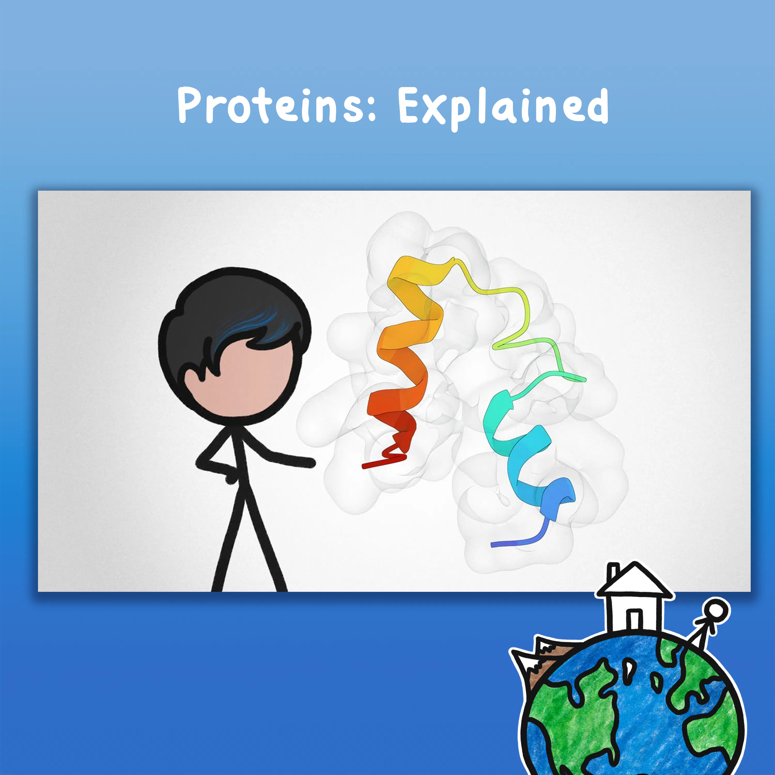
To learn more about this topic, start your googling with these keywords: - Amino acids: are organic compounds that contain amino (–NH2) and carboxyl (–COOH) functional groups, along with a side chain specific to each amino acid. - Proteins: are macromolecules composed of one or more long chains of amino acid residues. Most proteins fold into unique 3D structures. The shape into which a protein naturally folds is known as its native conformation. - Alpha helix (α-helix): is a common motif in the secondary structure of proteins and is a right hand-helix conformation in which every backbone N−H group hydrogen bonds to the backbone C=O group of the amino acid located four residues earlier along the protein sequence. - Beta sheet (β-sheet): is a common motif of the regular protein secondary structure and consists of beta strands (β-strands) connected laterally by at least two or three backbone hydrogen bonds, forming a generally twisted, pleated sheet. - Ribbon diagrams: are 3D schematic representations of protein structure that shows the overall path and organization of the protein backbone in 3D. Ribbon diagrams are generated by interpolating a smooth curve through the polypeptide backbone. α-helices are shown as coiled ribbons or thick tubes, β-strands as arrows, and non-repetitive coils or loops as lines or thin tubes. CREDITS ********* Ever Salazar | Co-Writer, Narrator, Illustrator and Director David Wych | Co-writer and Consultant Aldo de Vos, Know Art | Music MinuteEarth is produced by Neptune Studios LLC https://neptunestudios.info OUR STAFF ************ Sarah Berman • Arcadi Garcia Rius • David Goldenberg Julián Gustavo Gómez • Melissa Hayes • Alex Reich Henry Reich • Peter Reich • Leonardo Souza Ever Salazar • Kate Yoshida OUR LINKS ************ Youtube | https://youtube.com/MinuteEarth TikTok | https://tiktok.com/@minuteearth Twitter | https://twitter.com/MinuteEarth Instagram | https://instagram.com/minute_earth Facebook | https://facebook.com/Minuteearth Website | https://minuteearth.com Apple Podcasts| https://podcasts.apple.com/us/podcast/minuteearth/id649211176 OTHER CREDITS & REFERENCES ********************************** Goodsell, David (2006). Visual Methods from Atoms to Cells. Structure 13, Issue 3:347-354. doi:10.1016/j.str.2005.01.012 Protein 3D images created using Mol* (https://molstar.org/) and structure data from RCSB PDB (https://www.rcsb.org/) Mol* (D. Sehnal, A.S. Rose, J. Kovca, S.K. Burley, S. Velankar (2018) Mol*: Towards a common library and tools for web molecular graphics MolVA/EuroVis Proceedings. doi:10.2312/molva.20181103) Villin folding trajectory by Stefan Doerr - https://figshare.com/authors/Stefan_Doerr/748688 Clathrin Structure (PDB ID: 3IYV) Fotin, A., et al (2004). Molecular model for a complete clathrin lattice from electron cryomicroscopy. Nature 432: 573-579. doi:10.1038/nature03079 Immunoglobulin Structure (PDB IDs: 1IGT & 1IGY) Harris, L.J., et al (1998). Crystallographic structure of an intact IgG1 monoclonal antibody. J Mol Biol 275: 861-872. doi:10.1006/jmbi.1997.1508 ATP Synthase Structure (PDB IDs: 5ARE, 5ARI & 5FIL) Zhou, A., et al (2015). Structure and conformational states of the bovine mitochondrial ATP synthase by cryo-EM. ELife, 4. doi:10.7554/eLife.10180 RCSB PDB Molecule of the Month by David S. Goodsell (The Scripps Research Institute and the RCSB PDB) - https://pdb101.rcsb.org/motm/72 Photosystem II (PDB ID: 5XNL) Su, X., et al (2017). Structure and assembly mechanism of plant C2S2M2-type PSII-LHCII supercomplex. Science 357: 815-820. doi:10.1126/science.aan0327 Ribonuclease (PDB ID: 2AAS) Santoro, J., et al (1993). High-resolution three-dimensional structure of ribonuclease A in solution by nuclear magnetic resonance spectroscopy. J Mol Biol 229: 722-734. doi:10.1006/jmbi.1993.1075 Myosin (PDB ID: 1B7T) Houdusse, A., et al (1999). Atomic structure of scallop myosin subfragment S1 complexed with MgADP: a novel conformation of the myosin head. Cell 97: 459-470. doi:10.1016/s0092-8674(00)80756-4